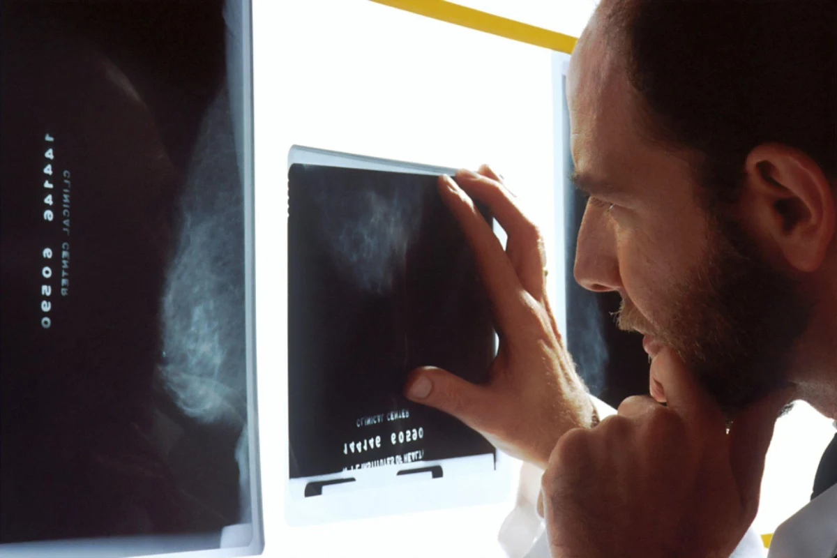
Diagnostic imaging supports preventive healthcare by letting providers see changes inside the body before they grow into bigger problems. A bone density scan can show early bone loss years before a fall turns into a fracture. An ultrasound can give a clear view of organs and blood vessels without any radiation at all. When that information arrives early, you and your provider have more time and more options to work with.
Most people only think about imaging after something starts to hurt. Many scans, however, are ordered as a quiet check on health that looks perfectly fine from the outside. If your provider suggests one, the process usually starts with a requisition form. For people in the Hamilton area, Alfa Scan Diagnostic Imaging Clinics offer X-ray, ultrasound and bone mineral density exams by appointment in Hamilton and Caledonia.
An imaging exam produces pictures of the bones and soft tissue inside your body. A radiologist then reviews those pictures and sends a written report to the provider who ordered the test. That report describes what the images show, such as the density of a bone or the size of an organ, so your provider can compare it with what they already know about you.
It helps to think of imaging as one important source of evidence that your provider weighs with everything else they know. They combine the findings with your symptoms and other test results to understand what may be going on. From there, they decide on the right next step together with you, and that combined view is what turns a picture into a useful plan.
Osteoporosis earns its reputation as a silent condition because bone loss usually causes no symptoms until a bone breaks. According to Osteoporosis Canada, at least one in three women and one in five men will have an osteoporotic fracture during their lifetime. The organization also estimates that more than 2.3 million Canadians are living with the condition.
The encouraging part is that bone density can be measured long before a fracture ever happens. A Public Health Agency of Canada report found that only 14.7% of people had a bone density test within a year after an osteoporosis-related fracture. That leaves plenty of room for earlier, more relaxed conversations about bone health.
The test itself is simple and comfortable for most people. MedlinePlus explains that a bone density scan uses very low doses of radiation to measure calcium and other minerals in your bones. You lie on your back on a padded table, and the test usually takes 10 to 30 minutes. Results are often given as a T-score, where -1.0 or higher is considered normal and -2.5 or lower points toward probable osteoporosis.
Ultrasound uses high-frequency sound waves to build live images of the inside of the body. The National Institute of Biomedical Imaging and Bioengineering notes that diagnostic ultrasound is generally regarded as safe and does not produce the ionizing radiation used in X-rays. That is one reason providers often choose it when they want a closer look at soft tissue.
Providers commonly use ultrasound to look at organs in the abdomen and pelvis as well as major blood vessels. One well-known preventive example comes from the Canadian Task Force on Preventive Health Care. In 2017, it recommended one-time ultrasound screening for abdominal aortic aneurysm in men aged 65 to 80, ideally after a conversation between each patient and their provider.
X-rays are one of the most familiar imaging tests, and they remain a quick way for providers to check bones and the chest. Beyond new injuries, they help providers follow how a fracture is healing. They also make it easy to compare current images with older ones to see whether anything has changed over time
Because X-rays use a small amount of radiation, providers order them when the information is likely to help with your care. If you are curious about why a particular X-ray is being recommended, your provider will be glad to explain. Knowing the reason often makes the appointment feel more familiar.
A little preparation makes the visit feel easier and helps the images come out clearly. Bring your requisition and health card, and wear comfortable clothing without metal zippers or buttons. It also helps to ask the clinic ahead of time whether your exam needs any preparation. Some abdominal or pelvic ultrasounds, for example, ask you to arrive with an empty stomach or a full bladder.
After the exam, the radiologist’s report goes to your provider, who will walk you through what it means and whether anything comes next. Many clinics now let patients see their own images and reports online. Alfa Scan, for example, uses PocketHealth so patients can securely view their images and reports and share them with other members of their care team.
The real value of preventive imaging comes from what you and your provider do with the information over the years. A baseline bone density result or a clear ultrasound becomes a reference point that makes future changes easier to spot. If imaging has never come up during a checkup, especially as you reach your fifties and sixties, it is a reasonable topic to raise. Your next appointment is a good moment to decide together what makes sense for your health.




Sign up to receive exclusive content updates, fashion & beauty tips!

Copyright © 2026 All Rights Reserved By Health Glowera