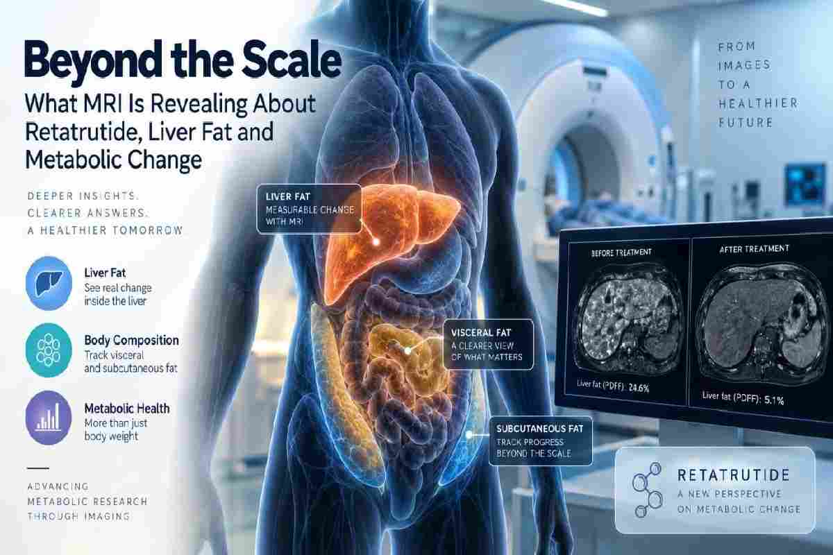
When metabolic drugs make headlines, the discussion usually comes down to one number: body weight. It is easy to measure, easy to understand and easy to compare across time. But weight alone says very little about where fat is stored, how much is present in the liver, or whether two people who lose the same number of pounds are undergoing the same metabolic changes internally.
That is one reason Retatrutide has become an interesting case for imaging-based metabolic research. Much of the attention around the molecule has focused on changes in body weight, while MRI data from its Phase 2 program offered a closer look at what was happening inside the liver and abdominal fat compartments. Instead of asking only how much weight changed, researchers could begin asking where that change was taking place.
Medical imaging is particularly useful for this kind of work because it can measure tissues that are largely invisible from the outside. MRI can quantify liver fat and distinguish between different abdominal adipose-tissue compartments, adding another layer of information to familiar measures such as body weight, BMI and waist circumference.
Body weight is useful precisely because it is simple. A scale produces a number that can be measured repeatedly over time, making it one of the easiest endpoints to follow in a clinical study. The problem is that the number combines many different tissues and tells researchers almost nothing about how those tissues are distributed.
Fat beneath the skin is not metabolically identical to visceral fat surrounding abdominal organs, and neither is the same as fat deposited inside the liver. Two people can have similar BMIs while carrying very different amounts of hepatic or visceral fat, which can translate into very different metabolic profiles.
That distinction becomes more important as modern metabolic therapies produce larger changes in body weight. If a participant loses 15 or 20 percent of starting weight, researchers naturally want to understand what happened beneath the headline number. Did liver fat fall substantially? Was there a large reduction in visceral adipose tissue? Did different fat compartments respond at roughly the same rate?
A scale cannot answer those questions. Imaging can.
The liver occupies a central position in metabolism. It helps regulate glucose availability, processes lipids and responds continuously to changing concentrations of insulin, glucagon and circulating nutrients. When excess fat accumulates within liver cells, it can become part of a broader pattern of metabolic dysfunction.
This accumulation is now commonly discussed in the context of metabolic dysfunction-associated steatotic liver disease, or MASLD. It often occurs alongside obesity, insulin resistance and type 2 diabetes, although body size by itself is not a reliable way to determine how much fat is actually stored in the liver.
That makes liver fat a useful example of why researchers need measurements beyond body weight. A person may lose a meaningful amount of weight without anyone being able to determine the exact change in hepatic fat simply by looking at them or measuring their waist.
MRI-PDFF — magnetic resonance imaging proton density fat fraction — offers a non-invasive way to quantify that change. Rather than classifying the liver only as fatty or non-fatty, researchers can estimate the proportion of liver tissue composed of fat and follow that measurement across multiple study visits.
Retatrutide is being investigated as a single molecule with agonist activity at the GIP, GLP-1 and glucagon receptors. The first two pathways are closely associated with nutrient-responsive signaling, while glucagon introduces effects related to hepatic metabolism, substrate utilization and energy regulation.
That receptor profile makes liver imaging particularly relevant. If several metabolic pathways are being influenced at once, changes in total weight may reveal only one part of the response. Researchers also want to know whether changes are appearing directly in metabolically important tissues.
A Phase 2a substudy provided an opportunity to examine that question. The analysis included participants from the larger Phase 2 obesity trial who had elevated liver fat at baseline. Investigators used MRI-PDFF to quantify hepatic fat and also assessed visceral and abdominal subcutaneous adipose tissue.
The study therefore produced something much richer than another weight-loss percentage. It offered a way to examine how internal fat stores were changing while the broader metabolic response was unfolding.
The liver-fat changes were substantial, particularly at the higher studied doses. By week 24, mean relative reductions in liver fat ranged from roughly 43% in the lowest-dose group to more than 80% in the higher-dose groups, while the placebo group showed essentially no meaningful reduction over the same period.
The effect remained evident through week 48. In the higher-dose groups, mean relative liver-fat reductions remained above 80%, and a large proportion of participants reached liver-fat levels below the threshold used in the study to define resolution of steatosis.
Those figures need context. The analysis came from a relatively small Phase 2 substudy, not from a dedicated large-scale liver-disease outcomes trial, and retatrutide is not an approved treatment for MASLD. Even so, the findings provide a useful demonstration of what quantitative imaging can add to metabolic research.
Without MRI, researchers would have known that participants lost weight. With MRI, they could also measure what was happening inside the liver.
Imagine two participants who each lose 15% of their starting body weight. From the perspective of the scale, their outcomes look identical. Internally, however, one participant might experience a particularly large reduction in liver and visceral fat, while the other could show a different pattern of tissue change.
That is one reason the imaging component of the retatrutide peptide research is worth examining separately from the headline weight-loss results. Investigators were not limited to a single measurement; they could look at liver fat, visceral adipose tissue and abdominal subcutaneous adipose tissue as distinct compartments.
Visceral and subcutaneous fat are often grouped together in everyday conversations about body fat, but they occupy different anatomical locations and have different metabolic characteristics. Visceral fat surrounds internal organs within the abdominal cavity, while subcutaneous fat is stored beneath the skin.
Separating those compartments gives researchers a clearer view of how the body is changing during treatment rather than treating every lost kilogram as biologically identical.
MRI is often associated primarily with diagnosis. A scan is ordered to identify an abnormality, characterize a lesion or examine anatomy that cannot be evaluated adequately from the outside. In clinical research, however, imaging can also be used as a repeatable quantitative measurement.
Liver-fat assessment is a good example. MRI-PDFF can provide a numerical value that is measured at baseline and then compared with later scans. When acquisition and analysis methods are standardized, researchers can track changes over time rather than relying only on visual interpretation.
That is particularly useful in multicenter clinical studies, where consistency matters. Different hospitals may use different scanners, and participants may return for imaging months apart. Researchers therefore need protocols that make measurements sufficiently comparable across sites and visits.
The goal is no longer simply to produce a good-looking image. The image itself becomes data.
The imaging findings also highlight an important point about metabolic research: many outcomes are related without being interchangeable.
Reductions in body weight can be associated with reductions in liver fat and visceral adipose tissue, but knowing that someone lost weight does not tell researchers exactly how much their hepatic fat fraction changed. The same is true of waist circumference, which provides useful information about abdominal size but cannot directly quantify the fat content of the liver.
This is why quantitative imaging can add value even when simpler clinical measurements are already available. Researchers do not need MRI because weight has become irrelevant; they need it because weight answers a different question.
Weight asks how much the body changed overall. MRI can help show where some of that change occurred.
MRI is not the only technology being used to study what lies beneath total body weight. Researchers can also use methods such as dual-energy X-ray absorptiometry, or DXA, to distinguish fat mass from lean mass.
That distinction matters because a 20-pound reduction in total body weight does not reveal how much of that change came from fat tissue versus lean tissue. As metabolic therapies become capable of producing larger reductions in total weight, understanding body composition becomes increasingly important.
The broader trend is toward combining several types of measurements. Body weight provides one layer of information, MRI provides another, body-composition analysis adds another, and laboratory biomarkers can show changes in glucose regulation, lipids, liver biology and other metabolic processes.
Together, these data give researchers a much more complete picture than any one measurement could provide on its own.
The liver-fat work is especially relevant because it connects a drug mechanism with an organ that plays a central role in energy metabolism. Glucagon receptor signaling is closely linked with hepatic processes, making liver outcomes a natural area of interest when studying a triple GIP/GLP-1/glucagon agonist.
That does not mean the liver-fat changes can simply be attributed to glucagon activity alone. Retatrutide activates three receptor systems simultaneously, and clinical studies cannot neatly divide an observed outcome into separate percentages contributed by each receptor.
Weight loss itself can also contribute substantially to reductions in liver fat. The meaningful question is therefore not whether one receptor caused one specific percentage change, but what the combined physiological response looks like when several metabolic pathways are influenced together.
Quantitative imaging helps researchers describe that response with far more precision.
The MRI substudy was conducted within retatrutide’s Phase 2 program, but the molecule has since progressed into much larger Phase 3 trials. In 2026, Lilly reported substantial weight reductions from TRIUMPH-1, including an average reduction of 28.3% at 80 weeks with the 12 mg dose under the study’s efficacy estimand.
Those headline figures have naturally attracted attention, but they also make tissue-level research increasingly relevant. When changes in total body weight become large, understanding what is happening to liver fat, visceral fat, lean tissue and other metabolic compartments becomes more—not less—important.
The broader retatrutide research peptide program now extends across obesity and several obesity-associated conditions. Retatrutide nevertheless remains investigational, and its safety and efficacy continue to be evaluated in clinical trials.
Phase 3 weight-loss figures and Phase 2 imaging findings therefore answer different questions. One describes the magnitude of a clinical outcome across a large population; the other helps researchers understand part of the biological change taking place beneath that outcome.
The larger lesson extends beyond retatrutide.
As metabolic therapies become more effective at changing body weight, clinical research may increasingly move toward understanding the quality and distribution of those changes. Losing weight matters, but researchers also want to know what happened to metabolically active tissues and organs along the way.
MRI is particularly well suited to this problem because it can repeatedly evaluate internal tissues without ionizing radiation. Depending on the protocol, it can help quantify hepatic fat, distinguish abdominal fat compartments and provide detailed anatomical information that would otherwise remain unavailable.
This matters because not every kilogram of body weight carries the same biological meaning. Changes in liver fat, visceral adipose tissue, subcutaneous fat and lean mass may each tell researchers something different about metabolic response.
The more precisely those changes can be measured, the easier it becomes to understand what a therapy is actually doing.
For years, one of the easiest ways to describe the effect of a metabolic intervention was to ask how much weight someone lost. That question remains important, but modern research increasingly has the tools to go further.
Researchers can now ask how liver fat changed, whether visceral adipose tissue declined, what happened to body composition and how those changes relate to glucose regulation and other metabolic outcomes. The retatrutide MRI substudy provides a useful example of how those additional measurements can alter the way a clinical trial is understood.
The scale tells researchers that the body changed.
MRI can help show where part of that change occurred.
That shift — from asking only “How much?” to also asking “Where?” — may become increasingly important as the next generation of metabolic research develops.
Retatrutide remains an investigational compound and is not currently approved by the FDA. The clinical findings discussed here come from controlled research studies and should not be interpreted as recommendations for use.




Sign up to receive exclusive content updates, fashion & beauty tips!

Copyright © 2026 All Rights Reserved By Health Glowera